Multifocus microscopy with high resolution
30.11.2022 - A new type of microscope can take 3D images of cells while working in a natural environment.
To observe living cells through a microscope, a sample is usually squeezed onto a glass slide. It then lies there calmly and the cells are observable. The disadvantage is that this limits how the cells behave and it only produces two-dimensional images. Researchers from UiT The Arctic University of Norway and the University Hospital of North Norway (UNN) have now developed what they are referring to as the next generation microscope. The new technology can take pictures of much larger samples than before, while living and working in a more natural environment.
-
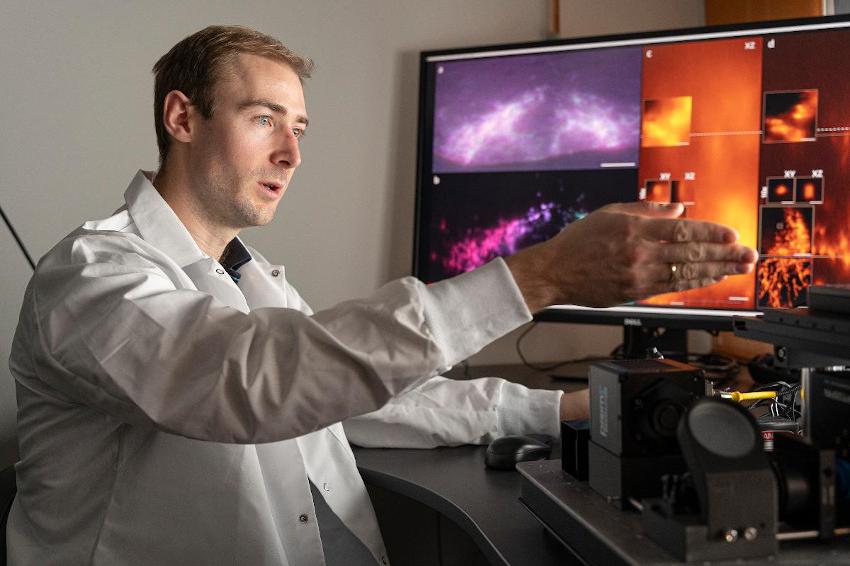 Florian Ströhl developes the next generation of microscopes. (Source: K. Rydland, UIT)
The technology provides 3D images where researchers can study the smallest details from several angles, clearly and visibly, sorted into different layers and all layers are in focus. 3D microscopes do already exist, but they work slowly and give poorer results. The most common type works by recording pixel after pixel in series, which are then assembled into a 3D image. This takes time and often they can't handle more than 1 to 5 shots a minute. It's not very practical if what you're going to photograph something that moves.
“With our technology, we can manage around 100 full frames per second. And we believe it is possible to increase this number. This is just what we have demonstrated with our prototype,” says Florian Ströhl, researcher at UiT. The new microscope is a multifocus microscope, which provides completely clear images, sorted into different layers, where you can study the cells from all angles. “It’s a big deal. The fact that we manage to get all this in one take, it is a huge development,” says Ströhl.
Ströhl explains that we are not talking about 3D in the form most of us know it. While in a traditional 3D image you will be able to perceive some kind of depth, with the new technology you are also be able to see behind objects. Ströhl uses an example where you see a jungle scene in 3D at the cinema. “In a normal 3D image, you can see that the forest has a depth, that some leaves and trees are closer than others. With the same technology used in our new 3D microscope, you are also able to see the tiger hiding behind the bushes. You are able to see and study several layers independently,” says Ströhl.
Ströhl has collaborated with researchers and doctors from the University Hospital of North Norway (UNN) in the development of this technology. Among other things, they work to understand and develop better treatment methods for various heart diseases. Studying a living human heart is challenging, both for technical reasons and not least for ethical reasons. Thus, researchers have used stem cells that are manipulated so that they mimic heart cells. In this way, they can grow organic tissue that behaves as it would in a human heart, and they can study and test this tissue to understand more about what is happening.
This tissue is almost like a small lump of live meat, about 1 centimeter in size. This makes for a very demanding test situation, where heart cells beat and are in constant motion along it the fact that the sample is too large to study with traditional microscopes. The new microscope handles this well. “You have this pumping lump of meat in a bowl, which you want to take microscope pictures of. You want to view at the very smallest parts of this, and you want super high resolution. We have achieved this with the new microscope,” says Ströhl.
Kenneth Bowitz Larsen heads a large laboratory with advanced microscopes that are used by all the research groups at the Faculty of Health at UiT. He has tested this new microscope, and is optimistic. “The concept is brilliant, the microscope they have built does things that the commercial systems do not,” Larsen explains. The laboratory he heads mainly uses commercial microscopes from different suppliers.
“Then we also collaborate with research groups like the one Florian Ströhl represents. They build microscopes and test optical concepts, they are in a way like the formula 1 division of microscopy,” Larsen says. Larsen has great faith in the new microscope Ströhl has created. The commercial microscopes must be usable for all kinds of possible samples, while the microscope Ströhl has developed is more tailored to a specific task.
“It is very photosensitive, and it can depict the specimen in various focuses. It can work its way through the sample and you can view both high and low. And it happens so fast that it can practically be seen in real time. It's an extremely fast microscope,” Larsen says. According to Larsen, the tests so far show that this works well, and he believes this type of microscope can eventually be used on all types of samples where you look at living things that move.
He also sees another advantage with the speed of this microscope. “Bright lights are not kind to cells. Since this microscope is so fast, it exposes the cells to much shorter illumination and is therefore more gentle,” he explains. The prototype of the microscope works and is operational. The researchers are currently working on creating an upgraded version that is easier to use, so that more people are able to operate and use the microscope.
The researchers have also applied for a patent and are also looking for industrial partners who will develop this into a microscope that will be available for sale. In the meantime, the prototype will be made available to local partners who can benefit from the new technology. “We will also offer it to others in Norway, if they have particularly demanding samples that they want examined,” says Ströhl. (Source: UiT)